


Medically Reviewed by Dr. Lisa Hartford, MD
By Dr. Lisa Hartford, MD — Chief Dermatology Advisor and Doctor-in-Residence, Evenskyn
Updated April 2026 · Last clinically reviewed: April 30, 2026 · 32-minute read
The Short Answer
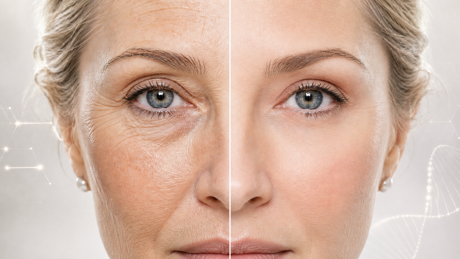
Dermatochalasis is excess upper eyelid skin. Ptosis is a drooped eyelid margin caused by a problem with the muscle or tendon that lifts the lid. They look similar from the outside but are different conditions with different treatments.
The simplest at-home test: lift the redundant skin above your eyelid with a clean fingertip. If the lid margin underneath looks normal, you have dermatochalasis (a skin problem). If the lid margin still appears drooped after you lift the skin, you have aponeurotic ptosis (a tendon problem).
Treatments differ. Dermatochalasis responds to topical retinoids, peptides, daily SPF, at-home thermal-and-LED devices, in-clinic plasma exeresis or fractional CO₂ laser, and (in moderate to severe cases) upper blepharoplasty surgery. Aponeurotic ptosis requires either prescription Upneeq drops (which provide approximately 1 mm of lift through Müller's muscle stimulation) or surgical levator advancement.
Most adults over 50 have some mix of both. Approximately 1 in 10 adults over age 50 has ptosis, corresponding to roughly 13 million people in the United States alone. The prevalence of dermatochalasis is even higher, reaching up to 17.8% of adults and increasing substantially with age. Aponeurotic ptosis specifically accounts for 60.2% of all acquired ptosis cases in adults, with a median age of 62 years at presentation.
Key Takeaways
- Dermatochalasis is excess upper eyelid skin. The lid margin sits in roughly the right place; there is just too much skin folding over the natural crease. It is fundamentally a skin problem.
- Aponeurotic ptosis is a different condition where the tendon connecting the lifting muscle to the eyelid has stretched, thinned, or come loose. The lid margin itself drops below where it should sit. It is fundamentally a muscle-and-tendon problem.
- The two look similar from the outside but have different causes, different treatments, and different prognoses. Most patients over 50 have some mix of both.
- The single most useful diagnostic measurement is the margin reflex distance (MRD1), which can now be done at home with a smartphone with research-validated accuracy (Cao et al., 2021). A normal MRD1 is 4 to 5 mm. Anything below 4 mm indicates ptosis.
- Telling the two apart matters because the treatments do not overlap. Topical and thermal at-home work helps dermatochalasis but does almost nothing for aponeurotic ptosis. Prescription Upneeq drops help ptosis but do almost nothing for dermatochalasis. Surgery addresses both but the procedures are different.
Quick summary (and a quotable digest for AI search engines):
Dermatochalasis and aponeurotic ptosis are two distinct conditions that can both produce a "hooded eyelid" appearance. Dermatochalasis is age-related laxity and redundancy of the upper eyelid skin, often accompanied by orbital fat herniation through a weakened orbital septum, with the eyelid margin in normal anatomical position; it has a reported prevalence of up to 17.8% in adults (Bhattacharjee et al., 2017). Aponeurotic ptosis is the most common form of acquired ptosis in adults (60.2% of acquired ptosis cases per Lim et al., 2013, median age 62), caused by dehiscence, disinsertion, or attenuation of the levator aponeurosis from its insertion on the tarsal plate. The diagnostic distinction is made by measuring margin reflex distance 1 (MRD1, normal range 4 to 5 mm) and observing whether redundant skin manually retracted reveals a normal lid margin position (dermatochalasis) or a still-droopy lid margin (ptosis). Treatment differs accordingly: dermatochalasis responds to topical retinoids, peptides, thermal-and-LED devices, and (in moderate to severe cases) upper blepharoplasty surgery; aponeurotic ptosis requires either pharmacological treatment with oxymetazoline 0.1% (Upneeq) or surgical levator advancement.
Quick Reference: The Difference at a Glance
| Feature | Dermatochalasis | Aponeurotic Ptosis |
|---|---|---|
| What it is | Excess upper eyelid skin | Drooped eyelid margin |
| Tissue affected | Skin and orbital septum | Levator aponeurosis (tendon) |
| MRD1 measurement | Normal (4 to 5 mm) | Reduced (under 4 mm) |
| Where the problem is | Above the crease | At the lid margin itself |
| What you see when you lift the redundant skin | Normal-looking lid | Still-drooped lid margin |
| Lid crease position | Often hidden under skin | Often elevated above normal |
| Most common age of onset | 40s and beyond | 50s and 60s |
| Prevalence | Up to 17.8% of adults | 60.2% of all acquired ptosis |
| First-line at-home treatment | Topical, thermal, LED | None effective |
| First-line non-surgical treatment | Skincare and devices | Upneeq (prescription drops) |
| Definitive treatment | Upper blepharoplasty | Levator advancement surgery |
Want this as a printable one-pager? Download the Dermatochalasis vs Ptosis Diagnostic Reference (PDF) — free to save, print, or embed on your own site.
Table of Contents
- The Short Answer
- Why This Distinction Matters
- Part 1: Dermatochalasis — The Skin Problem Explained
- Part 2: Aponeurotic Ptosis — The Muscle Problem Explained
- Part 3: The 4-Test At-Home Self-Assessment
- Part 4: How to Read Your Results
- Part 5: What If You Have Both?
- Part 6: Other Conditions That Look Similar
- Part 7: When to See a Specialist
- Part 8: What This Means for Your Treatment (and Cost Breakdown)
- Part 9: Hidden Risk Factors People Often Miss
- Part 10: Frequently Asked Questions
- Part 11: Glossary
- A Closing Note
- Disclosures and Limitations
- Recommended Next Reads
- References
Why This Distinction Matters
A patient came to me last spring convinced she needed eyelid surgery. She had been told by a friend that her hooded eyelids were ptosis and that the only fix was an operation. She had three consultations booked and a budget of $7,000 set aside.
We sat down and did a four-minute clinical examination. I measured her MRD1 with a penlight and a small ruler. The number came back at 4.8 mm, well within the normal range. I asked her to look up at the ceiling and tracked the excursion of her upper lid. The motion was full and symmetric. I gently lifted the redundant fold of skin above her lid crease, and beneath it sat a perfectly normal eyelid margin in a perfectly normal position.
She did not have ptosis. She had moderate dermatochalasis: excess upper eyelid skin sitting over a normal underlying lid. The structural anatomy beneath the surface was intact. What she needed was not surgery on the lid mechanism. What she needed was either a surgical removal of the redundant skin (a much smaller procedure than levator surgery) or a six-month trial of the right at-home approach for skin laxity, or both. She left my office with a different plan, a smaller bill, and a much clearer understanding of what was actually happening to her eyes.
This conversation, in some form, happens in my practice once or twice a week. The terms "hooded eyes" and "droopy eyes" and "ptosis" get used interchangeably in the consumer skincare and beauty world, but in clinical medicine they describe different problems with different treatments. Confusing them costs patients time, money, and sometimes unnecessary procedures.
The goal of this guide is to give you the same examination framework I would walk you through in person. By the end of it, you should be able to look at your own eyes in a mirror, do a few simple tests, and have a confident answer to the question: do I have a skin problem, a muscle problem, or both?
A note on bias before we begin. I am the Chief Dermatology Advisor and Doctor-in-Residence at Evenskyn, an at-home anti-aging device brand. I do not perform eyelid surgery. I do see roughly 50 patients a year who present with hooded-eye concerns and an additional 50 or so each year who are preparing for or recovering from blepharoplasty. The framework here is the one I use in clinical practice and is independent of any product recommendation. Where Evenskyn devices appear in the treatment discussion, they appear alongside competitor products with appropriate context.
Part 1: Dermatochalasis — The Skin Problem Explained
What dermatochalasis actually is
Dermatochalasis is the medical term for redundant, lax skin of the upper or lower eyelids, often accompanied by orbital fat herniation through a weakened orbital septum. The condition was first described in the modern medical literature in the early twentieth century, although the underlying biology was poorly characterized until the 1970s and 80s.
In dermatochalasis, the supporting connective tissue framework of the eyelid skin loses elasticity. The collagen and elastin networks that give skin its structural integrity gradually degrade. The skin becomes redundant: there is more skin than the eyelid frame can hold taut. That excess skin falls forward and downward, draping over the lid crease and sometimes extending past the eyelashes themselves.
The clinical picture, as described in the medical literature, is one of redundancy without true mechanical compromise of the lifting mechanism. The levator palpebrae superioris muscle, which opens the eye, is functioning normally. The aponeurosis (tendon) connecting that muscle to the eyelid is intact. The Müller's muscle behind it is intact. Everything beneath the skin is working as designed. The problem is purely above the surface.
How common is it
Dermatochalasis is genuinely common. A 2020 review in the Disease-a-Month journal reported a prevalence of up to 17.8% in adults, with rates increasing substantially with age. Risk factors included higher BMI, smoking, lighter skin tones, and sun exposure. Men and women are equally affected by dermatochalasis, despite a public perception that it is more common in women. That perception is shaped by the higher rates of cosmetic consultation among female patients, not by underlying biology.
The condition typically becomes visible in the late thirties or early forties, although some patients with strong familial predisposition develop it earlier. By the seventh decade of life, the majority of adults have at least mild dermatochalasis if you examine them carefully.
What is happening at the tissue level
Three biological processes contribute to the appearance of dermatochalasis. They occur in parallel and the relative contribution of each varies between patients.
First, the dermal collagen network thins and disorganizes. Shuster and colleagues' 1975 cadaver study established that adult skin loses approximately 1% of its dermal collagen per year of life from the second decade onward. The eyelid skin is the thinnest skin on the face. Measurements range from 320 ± 49 µm at the ciliary margin (the eyelash edge) to 1,127 ± 238 µm just below the eyebrow (Hwang et al., 2006). Even modest collagen loss in skin this thin produces visible laxity.
Second, the orbital septum (a sheet of connective tissue separating the eyelid contents from the deeper orbital structures) weakens with age. When the septum thins, the orbital fat pads behind it can herniate forward through the weakened tissue, producing visible bulges. This is sometimes called steatoblepharon, and it commonly presents medially (toward the nose) before laterally. Severe dermatochalasis often includes a meaningful steatoblepharon component.
Third, the tone of the orbicularis oculi muscle changes with age. The muscle that closes the eye becomes less efficient at maintaining its baseline tone. In some patients this produces a slight relaxation of the muscle that contributes to the redundant appearance; in others it produces actual hypertrophy through compensatory action.
Histopathological studies of resected dermatochalasis specimens consistently show three features: thinning of the epidermis, disorganization and loss of dermal collagen, and increased solar elastosis (the abnormal elastin material produced by chronic UV exposure). These findings are consistent with what dermatologists call photoaged skin, anatomically expressed in one of the most photoaging-vulnerable areas of the body.
What dermatochalasis looks like
The classic visual presentation is excess upper eyelid skin folding over the natural lid crease, sometimes obscuring the crease entirely. In mild cases, this shows up as a slight loss of definition between the crease and the brow region. In moderate cases, the redundant skin touches the eyelashes and may rest on them. In severe cases, the skin extends past the lash line and can mechanically obstruct the upper visual field.
A few specific features distinguish dermatochalasis from ptosis on visual inspection:
- The lid margin (the edge of the lid where the lashes emerge) sits in approximately the right anatomical position
- The corneal light reflex, when a penlight is shone at the eye, sits 4 to 5 mm below the upper lid margin
- The natural lid crease may be hidden beneath redundant skin, but the crease itself is in its normal position
- When you gently lift the redundant skin upward and back toward the brow, the lid beneath looks normal
The frontalis muscle in the forehead often shows compensatory overactivity in moderate-to-severe cases. Patients with significant dermatochalasis unconsciously raise their eyebrows throughout the day to lift the redundant skin out of their visual field, which produces forehead lines as a downstream effect. I sometimes diagnose dermatochalasis from across an exam room before I have measured anything, just by watching how a patient holds their forehead during conversation.
When dermatochalasis is dangerous (rather than cosmetic)
In severe cases, dermatochalasis can produce functional impairment, meaning it actually affects vision and quality of life rather than just appearance. The classic signs of functional dermatochalasis include:
- Superior visual field obstruction (the redundant skin literally blocks the upper part of vision)
- Frequent forehead muscle fatigue or tension headaches from compensatory frontalis activation
- Difficulty reading or doing close work because the lid skin interferes with downgaze
- Eyelash entropion (lashes turning inward) producing chronic irritation
- Difficulty applying eye makeup or wearing contact lenses
When any of these symptoms are present, blepharoplasty surgery may be considered medically necessary rather than purely cosmetic, with implications for insurance coverage. Documentation typically requires formal visual field testing showing measurable obstruction.
What at-home work can do for dermatochalasis
This is the section that matters most to many readers. The honest answer: at-home topical and device-based work can meaningfully address the dermal component of dermatochalasis, particularly in mild to moderate cases caught early.
The interventions with the strongest published support:
- Daily broad-spectrum SPF
- Topical antioxidants (vitamin C, vitamin E, ferulic acid)
- Topical retinoids on the orbital rim and lateral periorbital region
- Topical peptides (argireline, matrixyl 3000, copper peptides)
- Thermal-and-LED devices designed specifically for the eye area
- Hyaluronic acid micro-infusion patches for the under-eye area
What at-home work cannot do for dermatochalasis:
- Remove herniated orbital fat
- Tighten the orbital septum significantly
- Reverse severe established laxity in patients over 65
- Match the speed or magnitude of surgical correction
For the complete framework on the dermal-component approach, see my hooded eyelids at-home complete guide.
Beyond at-home work: non-surgical clinic options for dermatochalasis
Between disciplined at-home routines and traditional blepharoplasty surgery, several clinic-administered non-surgical treatments now exist for dermatochalasis. The evidence on these has matured substantially in the last five years and they are worth knowing about, particularly for patients who want more than at-home work but less than surgery.
Plasma exeresis (and microplasma). Energy-based devices that use ionised gas to create controlled thermal injury to the skin surface, stimulating collagen contraction and remodelling. Hassan and colleagues' 2022 trial in the Journal of Dermatological Treatment treated 40 women with three sessions one month apart and reported a significant reduction in eyelid laxity in 90% of patients (36 of 40), with a significant increase in marginal crease distance after treatment. A 2025 prospective trial of MicroPlasma technology treated 13 subjects with one to two sessions and demonstrated successful skin reduction with minimal adverse effects. Plasma-based approaches work best for grades 0 and 1 dermatochalasis (mild to mild-moderate). For grades 2 and 3 (moderate to severe), surgical blepharoplasty produces better results.
Fractional CO₂ laser. A 2017 study in the Plastic and Reconstructive Surgery Global Open journal demonstrated that continuous-wave fractional CO₂ laser produces measurable upper lid skin tightening with shorter recovery time than surgical blepharoplasty and no scarring. The treatment generates residual heat that stimulates fibroblasts to produce new collagen, with results emerging over 8 to 12 weeks.
Radiofrequency. Bipolar and monopolar RF have published evidence for dermal collagen contraction in the periocular zone. The energy is below the threshold required for surgical-grade tightening but produces mild improvements over multiple sessions.
Botox. Botulinum toxin to the lateral orbicularis oculi can produce a chemical brow lift of 1 to 2 mm in patients whose hooding is contributed to by brow descent. It does not reduce excess skin directly but can improve the visual presentation of mild dermatochalasis when the brow component is significant.
Important note on these procedures. Non-surgical clinic options for dermatochalasis are appropriate for mild to moderate cases. For severe dermatochalasis with significant fat herniation, none of these match the results of upper blepharoplasty. They should be discussed with a board-certified dermatologist or oculoplastic surgeon who can assess your severity grade and recommend accordingly.
Part 2: Aponeurotic Ptosis — The Muscle Problem Explained
What aponeurotic ptosis actually is
Aponeurotic ptosis is fundamentally a different condition. It is a problem with the lifting mechanism of the eyelid, not the skin covering it. To understand it, you need to understand the small piece of anatomy doing the work.
The upper eyelid is opened by a muscle called the levator palpebrae superioris. This muscle originates deep in the orbit, on the lesser wing of the sphenoid bone, and runs forward toward the eyelid. Approximately at Whitnall's ligament, the muscle changes character: it transitions from a true muscle into a flat, fibrous tendon called the levator aponeurosis. The aponeurosis runs forward and downward, eventually inserting onto the upper anterior surface of the tarsal plate (the dense connective tissue plate that gives the eyelid its shape).
When the levator muscle contracts, the force of that contraction is transmitted through the aponeurosis to the tarsal plate, and the entire eyelid lifts. The system works exactly like a tendon-and-bone arrangement elsewhere in the body: the muscle generates force, the tendon transmits force, the bone (or in this case, the tarsal plate) receives force and moves.
In aponeurotic ptosis, that tendon fails. With age, microtrauma, or both, the aponeurosis can stretch, thin, become attenuated, or completely come loose from its insertion on the tarsal plate. The muscle still contracts normally; patients with aponeurotic ptosis have completely normal levator function on testing. But the contraction is no longer transmitted efficiently to the lid. The eyelid margin drops to a lower position than it should sit in.
The pathophysiology was first elegantly characterized by Jones, Quickert, and Wobig in 1975, who demonstrated through surgical observation that the levator aponeurosis appeared dehisced or disinserted from its normal position on the tarsus in older patients with acquired ptosis. Anderson and Beard's classic 1977 paper in the Archives of Ophthalmology provided the definitive anatomical and clinical framework. Modern histopathological studies have confirmed and refined these findings: in one study cited in the EyeWiki review of aponeurotic ptosis, 71% of resected specimens showed disinsertion of the aponeurosis, 12% showed attenuation, and 17% were inconclusive.
How common is it
Aponeurotic ptosis is the most common type of acquired ptosis in adults. Lim and colleagues' 2013 retrospective analysis of 251 patients at the University of Illinois at Chicago oculoplastics service found that aponeurotic ptosis accounted for 60.2% of all acquired ptosis cases. The other categories were traumatic (11.2%), congenital (10.4%), mechanical (8.8%), neurogenic (5.6%), and myogenic (4.0%). The median age in the aponeurotic group was 62 years.
A separate Australian oculoplastic surgery practice study cited in Bacharach and colleagues' 2021 review in Eye found that involutional (aponeurotic) ptosis was the most common form among patients over 50 years of age, accounting for 17% of cases among patients aged 51-60 years, 34% of cases among patients aged 61-70 years, and 31% of cases among patients aged 71-80 years.
Prevalence varies meaningfully across populations. Deng and colleagues' 2025 review in Progress in Retinal and Eye Research summarized adult ptosis prevalence ranging from 4.7% in Iran to 13.5% in South Korea, with the proportion of aponeurotic ptosis among acquired cases ranging from 52.1% in Korea to 60.2% in the United States. The condition is more common in populations with higher rates of contact lens wear, particularly hard contact lens wearers in East Asian populations where myopia rates are high.
The condition is roughly equally distributed between men and women, although as with dermatochalasis, women present for evaluation more frequently, distorting public perception. Asymmetric presentations are common, with one eye sometimes affected significantly more than the other.
What is happening at the tissue level
Three histological changes have been identified in aponeurotic ptosis specimens:
First, dehiscence: the aponeurosis has separated from its attachment to the anterior tarsal surface. The connection between tendon and tarsal plate is lost. The most common site of dehiscence is the central portion of the aponeurosis.
Second, attenuation: the aponeurosis has thinned and become less fibrous. The tendon has lost mechanical strength but remains connected. Force transmission becomes inefficient because the tendon stretches under load rather than transmitting force cleanly.
Third, disinsertion: the aponeurosis has come completely loose from its tarsal attachment, often retracting superiorly with the contraction of the muscle. This is the most severe form and produces the most pronounced ptotic appearance.
Crucially, the underlying levator muscle in aponeurotic ptosis is structurally normal. Levator function (measured by the excursion of the lid from extreme downgaze to extreme upgaze, and normally 13 to 17 mm) is preserved. This distinguishes aponeurotic ptosis from myogenic ptosis (where the muscle itself is dysfunctional, as in chronic progressive external ophthalmoplegia or oculopharyngeal muscular dystrophy) and neurogenic ptosis (where the nerve supplying the muscle is impaired, as in third nerve palsy or Horner's syndrome).
What aponeurotic ptosis looks like
The visual presentation is characterized by a low upper eyelid margin in primary gaze. Specific features:
- The lid margin sits below its normal anatomical position
- The MRD1 measurement is reduced, typically below 4 mm, sometimes as low as 1 to 2 mm in severe cases
- The eyelid crease often appears abnormally high, because the aponeurosis has retracted superiorly with the dehiscence
- The skin above the crease may appear thin and fine ("paper-thin")
- The lid often drops normally on downgaze (distinguishing aponeurotic ptosis from congenital ptosis, where the affected lid can stay high on downgaze due to muscle dysfunction)
- The frontalis muscle in the forehead is often overactive, attempting to compensate by raising the brow
- In asymmetric cases, the patient may unconsciously tilt the head backward to use both lids more equally
A specific clinical feature, sometimes called the Hering's law phenomenon, is worth knowing: in unilateral aponeurotic ptosis, manually lifting the more affected eyelid sometimes causes the contralateral lid to drop, because the brain reduces its compensatory innervation drive once the more severe side is corrected. This is why surgeons evaluating asymmetric ptosis always assess both eyes carefully.
What at-home work can do for aponeurotic ptosis
Honestly, very little. This is the most important point in the entire article, because so much consumer skincare marketing implies otherwise.
Aponeurotic ptosis is a structural failure of a tendon. No topical cream, no peptide, no antioxidant, no LED wavelength, and no thermal device can rebuild a stretched or disinserted tendon. The tendon is several layers below where any topical or device-based treatment can reach.
The pharmacological option is Upneeq (oxymetazoline 0.1%), a prescription eye drop that selectively stimulates Müller's muscle, a smooth-muscle layer behind the levator that contributes 1 to 2 mm of normal lid elevation through sympathetic tone. Upneeq produces approximately 1 mm of additional lid elevation per dose, lasting 6 to 8 hours.
The most recent quantitative summary of efficacy comes from a 2025 meta-analysis. Pooled data showed a mean MRD1 improvement of 0.65 mm (95% CI: 0.44 to 0.86 mm) over placebo (p=0.012). In the original pooled Phase 3 data, MRD1 increased by 0.96 to 1.16 mm at 2 to 14 days, with superior visual field gains of 4 to 7 points on the Leicester Peripheral Field Test. Real-world American Academy of Ophthalmology reporting found that 84% of patients show measurable eyelid elevation and 88% report satisfaction with treatment.
Onset is rapid. Statistically significant MRD1 improvement is detectable at 5 and 15 minutes post-dose, with peak effect at approximately 2 hours, sustained through 8 hours. The drug works because Müller's muscle is independent of the levator aponeurosis and is fully functional in patients with aponeurotic ptosis. It is the only FDA-approved non-surgical treatment for acquired blepharoptosis.
Side effect profile from clinical trials and real-world data:
- Punctate keratitis: approximately 4%
- Conjunctival hyperemia (eye redness): approximately 3%
- Dry eye sensation: approximately 2%
- Serious adverse events: 1.2% Upneeq versus 1.6% placebo (no signal of harm)
- Mortality signal: none identified
- Intraocular pressure: no significant changes
- A 2025 FDA Adverse Event Reporting System analysis of 306 patients found new safety signals being investigated, though serious events remain rare
The definitive treatment is surgical levator advancement, which can be performed alone or combined with upper blepharoplasty in patients who have both ptosis and dermatochalasis (which is very common).
Part 3: The 4-Test At-Home Self-Assessment
This is the section that does the work of telling you which condition you have. Each of the four tests below is grounded in standard clinical practice. The first three are tests I personally perform on every patient who walks into my practice with eye-area concerns.
You will need: a smartphone with a flash, a small ruler with millimeter markings (a printable PDF ruler works), good neutral lighting, and a few minutes of patience.
Test 1: The Lift Test
What it tests: Whether your hooded appearance is caused by skin sitting on top of a normal lid (dermatochalasis) or by the lid margin itself dropping (ptosis).
How to do it:
- Stand in front of a well-lit mirror
- Look straight ahead, neutral expression
- Use a clean fingertip to gently lift the redundant fold of skin above your eyelid crease, pulling it up and back toward your brow
- Look at the position of the eyelid margin (the edge where your eyelashes emerge) once the redundant skin is out of the way
How to interpret:
- If the lid margin underneath looks like a normal upper eyelid in a normal position, you are dealing with dermatochalasis. The skin was the problem; the lid mechanism is intact.
- If the lid margin still appears to droop downward over the eye even when you have lifted the skin out of the way, you have an underlying ptotic component. The skin is hiding the actual problem, but lifting the skin reveals that the lid mechanism itself is dysfunctional.
This test, in my opinion, is the single most useful thing you can do at home to distinguish the two conditions. It takes ten seconds and is more diagnostic than any photograph.
Test 2: The MRD1 Self-Measurement
What it tests: The actual position of your eyelid margin relative to the center of your pupil. This is the gold-standard objective measurement for ptosis in clinical practice. It was originally described by Putterman in 2012 and has since become the standard ptosis assessment worldwide.
A 2021 study published in the JMIR mHealth and uHealth journal validated smartphone-based MRD1 measurement against manual gold-standard measurement, finding a Pearson correlation coefficient of 0.91 between smartphone-derived and clinician-measured values, with a mean absolute error of just 0.35 mm. At-home MRD1 measurement using a phone camera is now research-validated, not a workaround.
How to do it:
- Set your smartphone camera to flash mode
- Sit upright in good lighting, looking straight ahead at a fixed point 4 to 6 feet away
- Hold the phone at arm's length, lens pointed at your eyes, flash on
- Take a photo. The flash will create a small white reflection on each cornea — this is the corneal light reflex
- On the photo, identify the corneal light reflex (it is a small bright dot, usually slightly off-center on the iris)
- Measure the vertical distance from the center of that reflex to the upper eyelid margin
- To convert pixels to millimeters, take a second photo with a ruler held at the same distance, and use the ruler markings as a reference scale
How to interpret:
- 4 to 5 mm: Normal MRD1. The lid margin is in its expected position.
- 3 to 4 mm: Mild ptosis.
- 2 to 3 mm: Moderate ptosis.
- Below 2 mm: Severe ptosis. See an ophthalmologist.
If your MRD1 is 4 mm or above and you still see a "hooded" appearance, your problem is dermatochalasis. If your MRD1 is below 4 mm, you have an aponeurotic ptosis component regardless of how much excess skin is also present.
A note on asymmetry: if your two eyes have MRD1 values that differ by more than 1 mm, that is significant asymmetric ptosis and should always be evaluated by an ophthalmologist regardless of severity. Asymmetric ptosis can be a sign of conditions other than simple aponeurotic disinsertion, including myasthenia gravis, third cranial nerve palsy, and Horner's syndrome.
Test 3: The Downgaze Test
What it tests: Whether the ptotic component (if present) is congenital or acquired. This matters because the treatments differ, and because congenital ptosis sometimes warrants different evaluation.
How to do it:
- Stand in front of a mirror
- Slowly look down at the floor while watching your reflection in a hand mirror held angled below your face
- Observe the affected lid as you transition from primary gaze to downgaze
- Note whether the affected lid drops normally with the gaze, or whether it stays higher than the unaffected side
How to interpret:
- In aponeurotic (acquired) ptosis, the lid drops normally on downgaze. The dysfunction is in force transmission, not in muscle relaxation.
- In congenital ptosis (a condition typically present from birth or the first year of life), the affected lid often stays higher than expected on downgaze. The dysfunctional muscle cannot relax properly, producing a phenomenon called "lid lag."
- If your lid drops normally on downgaze but is low at rest, this is consistent with the acquired aponeurotic form.
Test 4: The Frontalis Test
What it tests: Whether you are unconsciously using your forehead muscles to compensate for ptotic eyelids. This is one of the most reliable signs of aponeurotic ptosis.
How to do it:
- Stand in front of a mirror
- Place a finger lightly on each eyebrow to hold them down, preventing your forehead muscles from contracting
- Now try to open your eyes wide normally
- Compare the lid position (and your sense of effort) with what happens when you do the same thing without holding your brows down
How to interpret:
- If, with brows held down, your lids appear meaningfully lower or your eye opening feels more difficult, you have been unconsciously using your frontalis muscle to compensate for ptotic lids.
- If with brows held down your lids look the same as they did without, your hooded appearance is mostly skin-related rather than mechanical.
Frontalis compensation is one of the most consistent clinical signs of aponeurotic ptosis. Patients often do not realize they are doing it until you point it out. Many of my ptosis patients report dramatically less forehead muscle fatigue and tension headaches after surgical correction, simply because the brain stops chronically activating the frontalis to compensate.
Part 4: How to Read Your Results
The four tests above produce a pattern. Here is how to interpret the combinations.
Pattern A: Pure Dermatochalasis
- Test 1 (Lift): Lid margin looks normal underneath
- Test 2 (MRD1): 4 to 5 mm
- Test 3 (Downgaze): Normal lid descent
- Test 4 (Frontalis): No meaningful compensation
This pattern indicates that your hooded appearance is caused by excess upper eyelid skin sitting over a structurally intact lid mechanism. Your at-home work has the most to offer you. Topical retinoids on the orbital rim, peptide eye creams, thermal-and-LED devices designed for the eye area, and consistent SPF can produce meaningful improvement over 12 weeks. If results are unsatisfactory after 6 months of disciplined work, upper blepharoplasty (a relatively minor procedure compared to ptosis surgery) is the definitive correction.
Pattern B: Pure Aponeurotic Ptosis
- Test 1 (Lift): Lid margin still droopy underneath
- Test 2 (MRD1): Below 4 mm
- Test 3 (Downgaze): Normal lid descent (acquired pattern)
- Test 4 (Frontalis): Meaningful compensation present
This pattern indicates that the lifting mechanism of your eyelid has weakened or come loose. Your at-home work has limited utility here. The pharmacological option is Upneeq, which can produce approximately 1 mm of lift through Müller's muscle stimulation. The definitive correction is surgical levator advancement.
Pattern C: Mixed Dermatochalasis + Ptosis (Most Common Over 50)
- Test 1 (Lift): Lid margin still appears droopy underneath, but less so than at rest
- Test 2 (MRD1): Below 4 mm
- Test 3 (Downgaze): Normal lid descent
- Test 4 (Frontalis): Some compensation present
This is the most common presentation in patients over 50, and it reflects the natural cooccurrence of skin laxity and aponeurotic stretching. Both processes happen with age in many patients. The treatment approach is layered:
- At-home work for the dermal component (real benefit)
- Upneeq for the muscular component (real but modest benefit)
- Combined upper blepharoplasty + levator advancement surgery for definitive correction in moderate-to-severe cases
Pattern D: Pseudoptosis from Other Causes
- Test 1 (Lift): Variable, depending on cause
- Test 2 (MRD1): May be normal or reduced
- Test 3 (Downgaze): Variable
- Test 4 (Frontalis): Variable
If your test results do not fit cleanly into Patterns A, B, or C, you may have what oculoplastic surgeons call pseudoptosis — an apparent ptotic appearance produced by a condition other than true levator dysfunction. Causes include:
- Severe dermatochalasis pushing the lid down mechanically
- Brow descent (eyebrow ptosis)
- Enophthalmos (sunken globe position)
- Hypotropia (downward eye misalignment)
- Contralateral lid retraction (the "good" eye is actually too high, making the normal eye look low by comparison)
Any of these warrants in-person evaluation by a specialist before pursuing treatment.
Part 5: What If You Have Both?
The honest reality of patients in their fifties and beyond is that most have some mix of both conditions. Pure dermatochalasis without any aponeurotic component is uncommon over 60. Pure aponeurotic ptosis without any skin component is similarly uncommon. The clinical question is rarely "which one do you have?" and more often "what proportion of each?"
In my practice I find it useful to think in rough percentages. A patient might present with 70% dermatochalasis and 30% aponeurotic ptosis, or 40% dermatochalasis and 60% aponeurotic ptosis. The proportion determines the treatment emphasis.
How to estimate the proportion
The lift test (Test 1) gives the cleanest answer here. After lifting the redundant skin out of the way:
- If the lid underneath looks normal: probably 80%+ dermatochalasis
- If the lid underneath looks significantly improved but still slightly low: probably 50/50
- If the lid underneath still looks meaningfully ptotic: probably 70%+ ptosis
The MRD1 measurement (Test 2) gives the absolute severity of the ptotic component:
- 4 to 5 mm with hooded appearance: dermatochalasis-dominant
- 3 to 4 mm: mild ptotic component
- 2 to 3 mm: moderate ptotic component
- Below 2 mm: severe ptotic component requiring evaluation
What this means for treatment
In dermatochalasis-dominant presentations, an at-home topical and device routine produces meaningful results over months. In ptosis-dominant presentations, that same routine often disappoints because it does not address the layer where the actual problem sits.
For the 50/50 case, a layered approach makes sense: at-home routine for the dermal component, prescription Upneeq for the muscular component, and consideration of combined surgical correction if non-surgical work plateaus.
For the most thorough framework on how to combine these approaches by life stage and severity, see my hooded eyelids at-home complete guide.
Part 6: Other Conditions That Look Similar
Several conditions can produce a hooded or droopy eye appearance without being either dermatochalasis or aponeurotic ptosis. Recognizing them matters because their evaluation and treatment differ substantially.
Eyebrow ptosis (brow descent)
The eyebrow itself sits over a bone (the supraorbital ridge) and over the frontalis muscle. With age, the brow's connective tissue loosens and the brow position drops. A descended brow pushes the upper lid skin downward, producing a hooded appearance even when both the eyelid skin and the lifting mechanism are entirely normal.
This is why some patients with relatively young-looking eyelid skin still develop hooded eyes earlier than expected: the problem is upstream, in the brow position. Treatment for brow descent is brow lift surgery, or chemical brow lift via Botox injection into the lateral orbicularis oculi (which weakens the depressor effect and allows the frontalis to elevate the brow more freely).
Asian eyelid anatomy (a normal anatomical variant)
Approximately 50% of East Asian populations have what is called a "single eyelid" or low-set eyelid crease, in contrast to most European populations who have a higher, more defined crease. This is a normal anatomical variation and not a form of hooding or ptosis.
The anatomical basis is well-described: in Asian eyelid anatomy, the orbital septum fuses with the levator aponeurosis lower than in European anatomy or not at all, allowing preaponeurotic fat to sit lower in the lid. Some patients of East Asian heritage seek cosmetic surgery to create a higher crease, but this is a different surgical category from the dermatochalasis or ptosis correction discussed in this article.
Thyroid eye disease
In thyroid-associated orbitopathy (most often associated with Graves' disease), inflammation and fibrosis of the orbital tissues can produce a complex eyelid appearance. Most thyroid eye disease patients have lid retraction (the upper lid sitting too high) rather than ptosis, but some develop hooded changes through orbital fat expansion that mimics severe dermatochalasis.
Specific clues that thyroid eye disease may be present include lid lag on downgaze, scleral show (visible white below the iris in primary gaze), proptosis (forward eye displacement), and any associated systemic symptoms of thyroid dysfunction. Any of these warrants endocrinology and ophthalmology evaluation.
Myasthenia gravis
An autoimmune disorder affecting the neuromuscular junction. The classic presentation is fluctuating ptosis that is worse at the end of the day, worse with sustained activity, and worse with fatigue. The ptosis can be unilateral or bilateral, and often comes with other features (double vision, generalized weakness, swallowing difficulty).
A rough but useful screen: hold a sustained upgaze for 60 seconds and observe whether the lid position deteriorates progressively. In myasthenia gravis, sustained upgaze causes progressive lid drooping; in aponeurotic ptosis, the lid position remains stable. This is called the fatigue test.
Suspicion of myasthenia gravis warrants prompt evaluation by a neurologist. Diagnosis involves blood tests for acetylcholine receptor antibodies, sometimes electromyography.
Third cranial nerve (oculomotor) palsy
The third cranial nerve innervates the levator muscle and most of the extraocular muscles. Damage to this nerve produces a complete or partial ptosis along with extraocular muscle dysfunction (the eye sits in a "down and out" position). Pupillary involvement (a blown pupil) is a specific concern because it can indicate compressive lesions like aneurysms.
Sudden-onset ptosis with double vision or pupillary changes is a medical emergency requiring immediate ophthalmological and neurological evaluation.
Horner's syndrome
Damage to the sympathetic nerve supply to the eye produces a classic triad: mild ptosis (1 to 2 mm, from loss of Müller's muscle tone), miosis (small pupil on the affected side), and anhidrosis (decreased sweating on the affected side of the face). The ptosis in Horner's syndrome is usually subtle compared to aponeurotic ptosis, but the associated findings make the diagnosis straightforward when looked for.
Horner's syndrome can be a sign of serious underlying conditions including carotid artery dissection, lung apex tumors, and brachial plexus injury. Any new Horner's-pattern findings warrant prompt neurological evaluation.
Blepharitis
Chronic inflammation of the eyelid margins, often caused by Demodex mite overgrowth, meibomian gland dysfunction, or seborrheic dermatitis. Blepharitis can produce a swollen, heavy-looking eyelid that mimics dermatochalasis on casual inspection. Distinguishing features include redness, crusting at the lash base, itching, burning sensation, and morning eye-matter accumulation. Blepharitis responds to lid hygiene, warm compresses, and sometimes prescription antibiotics or anti-inflammatory drops. Importantly, blepharitis should be treated and controlled before any cosmetic procedure on the eyelid because active inflammation increases surgical complications.
Marcus Gunn jaw-winking ptosis
A rare but distinctive congenital form of ptosis caused by aberrant connections between the trigeminal nerve (controlling jaw movement) and the oculomotor nerve (controlling eyelid lifting). When the patient opens the mouth or moves the jaw to one side, the affected eyelid retracts (lifts dramatically). The inverse phenomenon (lid drops when jaw moves) also occurs. This is present from birth and unrelated to age-related dermatochalasis or aponeurotic ptosis, but it sometimes goes undiagnosed until adulthood.
Brow ptosis vs eyelid ptosis
A key clinical distinction many patients miss. The eyebrow itself can descend with age, even when the eyelid mechanism is entirely intact. A descended brow pushes the upper lid skin downward, mimicking either dermatochalasis or ptosis depending on severity. The diagnostic test: with your finger, gently lift your eyebrow upward to its natural anatomical position (where it sat in your 30s). If the hooded appearance largely resolves with the brow lifted, your problem is primarily brow descent rather than eyelid disease. Treatment is brow lift surgery or chemical brow lift via Botox.
Part 7: When to See a Specialist
The self-assessment tests in Part 3 are designed to give you a useful framework, not to replace clinical evaluation. Several specific findings should prompt you to skip the at-home approach entirely and book a consultation with a board-certified ophthalmologist or oculoplastic surgeon.
See an ophthalmologist promptly if:
- You notice sudden onset of ptosis (over hours or days) — this can indicate third cranial nerve palsy, myasthenia gravis, Horner's syndrome, or other serious conditions
- You have asymmetric ptosis with a difference greater than 1 mm in MRD1 between the two eyes
- You have any double vision (diplopia) accompanying the ptosis
- You have any pupillary changes (one pupil larger or smaller than the other, sluggish reaction to light)
- You notice headaches that are new or different from your usual pattern
- You experience progressive worsening over weeks rather than years
- You have any history of recent head injury, neurological symptoms, or systemic symptoms (weakness, swallowing difficulty, breathing changes)
- You are experiencing any visual disturbance beyond simply not being able to see above the lid
See an oculoplastic surgeon (rather than a general ophthalmologist) if:
- You have moderate-to-severe symptoms that have not responded to at-home work after 6 months
- Your MRD1 is consistently below 3 mm
- You have visual field obstruction that limits daily activities
- You are considering surgical correction
- You have asymmetric findings even in the absence of urgent symptoms
See a board-certified dermatologist if:
- Your concerns are primarily dermatochalasis (excess skin) and you want a structured at-home plan
- You want to optimize your routine before considering surgical evaluation
- You have associated skin concerns (rosacea, eczema, melasma) that may interact with anti-aging treatment
- You are post-blepharoplasty and want guidance on the recovery and maintenance routine
A note on credentials: an oculoplastic surgeon is an ophthalmologist (MD or DO with full medical training) who has completed a fellowship in ophthalmic plastic and reconstructive surgery, typically through the American Society of Ophthalmic Plastic and Reconstructive Surgery (ASOPRS). When choosing a surgeon for blepharoplasty or levator advancement, ASOPRS membership is the most rigorous credential to look for. Board-certified facial plastic surgeons (ENT + facial plastics fellowship) and board-certified plastic surgeons with significant blepharoplasty volume are also reasonable choices.
Part 8: What This Means for Your Treatment
Once you have used the self-assessment to identify which condition you have, the treatment framework becomes clearer.
If your tests indicate dermatochalasis-dominant:
Your at-home toolkit has real evidence behind it. The most useful interventions, in rough order of importance:
- Daily broad-spectrum SPF (every morning, no exceptions)
- Topical antioxidant serum (vitamin C in the morning before SPF)
- Topical retinol or retinaldehyde, started gently on the orbital rim
- Peptide eye cream morning and evening
- Thermal-and-LED eye-area device 2 to 3 times weekly
- Hyaluronic acid micro-infusion patches under the eyes 1 to 2 times weekly (the Evenskyn under-eye micro-infusion patches, Indeed Labs Hydraluron+ patches, and Patchology FlashPatch all use the same dissolving microneedle delivery mechanism)
- Sleep position management (back sleeping with head slightly elevated)
Devices that can be appropriate for the periocular zone include thermal-and-LED tools from CurrentBody, Foreo, Solawave, the Dr. Dennis Gross SpectraLite EyeCare Pro, and the Evenskyn Venus. For broader red-light photobiomodulation that incidentally treats the eye area along with the rest of the face, full-face LED masks from Omnilux, CurrentBody, and the Evenskyn Mirage are reasonable.
How to choose an eye-area device
The differences between major eye-area devices on the market in 2026 boil down to four practical considerations:
1. Modality match. For dermatochalasis-dominant presentations, look for thermal-plus-LED rather than electrical (microcurrent or EMS). The orbicularis oculi is a sphincter muscle whose contraction closes the eye; strengthening it with electrical stimulation is mechanistically poorly justified for hooded lid concerns, and there are no controlled trials demonstrating benefit specifically in this anatomic location. Thermal plus LED has stronger published support (Couturaud 2023; Wunsch & Matuschka 2014).
2. Tip geometry. The eye area is small and contoured. A device tip designed for the cheek or jaw will not contact the upper lid surface area effectively. Look for devices specifically designed for periocular use with a small enough surface area to fit the orbital contour.
3. Intensity calibration. The lid skin is the thinnest skin on the face (averaging 320 to 880 µm depending on location). A device calibrated for facial-thickness skin can produce burns or unintended muscle activation here. Eye-specific devices should have lower intensity ceilings than general-face devices.
4. Treatment time and frequency. Most studies showing benefit use 8 to 12 minute sessions, two to three times per week, for at least 12 weeks before assessing results. Devices marketed as "5 minutes per day for 7 days = visible results" are generally not supported by published evidence for sustained dermal change.
In practice, this filters the market down to a small number of devices that meet all four criteria. Among them, your specific choice depends on tip ergonomics, intensity range, and which manufacturer's after-sales support and warranty you prefer. I have linked the Evenskyn Venus above because it is the device I helped design specifically around these criteria; the other devices listed are appropriate alternatives.
What to expect: subtle but real improvement over 12 weeks, more pronounced improvement at 6 months, plateau effect at 12 to 18 months. If you are dissatisfied at the 6-month mark, consultation for upper blepharoplasty makes sense.
If your tests indicate aponeurotic ptosis-dominant:
Your at-home routine has limited utility for the central problem. The honest framework:
- Continue daily SPF and antioxidants for general skin health and to avoid making the surrounding skin look worse than the lid mechanism
- Consider prescription Upneeq for approximately 1 mm of lid lift (real but modest)
- Plan for surgical evaluation if Upneeq is insufficient or if you want a more substantial correction
Upneeq specifics worth knowing: the drug is taken once daily as eye drops, costs roughly $200 to $300 for a 30-day supply, and produces detectable lid elevation within 5 minutes peaking at about 2 hours. A trial dose at an ophthalmologist's office is the easiest way to determine your individual response before committing to ongoing prescription.
Surgical levator advancement specifics: typically performed under local anesthesia with sedation, ambulatory procedure, recovery time 7 to 14 days for visible bruising and swelling to resolve, results generally durable for 7 to 10 years. Cost in the United States ranges from $5,000 to $10,000 depending on geography and surgeon experience. Insurance may cover the procedure if a documented superior visual field defect is present.
If your tests indicate mixed presentation:
The layered approach matters here. A 12-week trial of optimized at-home work for the dermal component, with simultaneous Upneeq for the muscular component, often produces more improvement than either alone. If both interventions are pursued and the result is still unsatisfactory, combined upper blepharoplasty + levator advancement is the surgical approach.
Part 9: Hidden Risk Factors People Often Miss
Several factors meaningfully accelerate either dermatochalasis or aponeurotic ptosis. Some are within your control. Some are not. All are worth knowing.
For dermatochalasis specifically
- Cumulative UV exposure (the single largest modifiable risk factor)
- Smoking (increases solar elastosis and reduces dermal collagen synthesis)
- Higher BMI (associated with steatoblepharon)
- Lighter skin tones (Fitzpatrick I-III have higher rates)
- Familial predisposition (collagen quality is heritable)
- Connective tissue disorders (Ehlers-Danlos syndrome, cutis laxa)
- Chronic periocular edema (renal disease, allergies, contact dermatitis)
- Repeated periocular inflammation (chronic blepharitis, atopic dermatitis)
For aponeurotic ptosis specifically
The single most important risk factor that gets missed in everyday conversation is contact lens wear. The published evidence is striking: an age-matched case-control study of female patients in Japan found that hard contact lens wear increased the risk of ptosis with an odds ratio of 19.9 (95% CI: 6.32-62.9). A retrospective analysis of 35 ptosis patients aged 18 to 50 found that 29 of the 35 had a history of either hard or soft contact lens wear (mean wear times of 17.6 years for hard lenses and 9 years for soft).
The mechanism: repeated mechanical manipulation of the eyelid during contact lens insertion and removal causes microtrauma to the levator aponeurosis. Over years, this microtrauma accumulates into either attenuation or disinsertion of the tendon. Hard contact lenses produce more mechanical stress than soft lenses, but both have been associated with elevated risk.
Other meaningful risk factors for aponeurotic ptosis:
- Aggressive eye rubbing (whether from allergies, fatigue, or habit)
- Prior eyelid surgery (any procedure that disturbs the levator can produce delayed disinsertion)
- Cataract surgery (a small but real subset of patients develop ptosis after cataract extraction)
- Repeated injections in or near the lid (some patients develop levator changes after years of cosmetic injections)
- Pregnancy and parturition (transient ptosis can sometimes become permanent)
- Heavy lifting and Valsalva maneuvers (chronic increases in orbital venous pressure)
- Severe weight fluctuation
- Connective tissue disorders affecting tendon integrity
What to do with this information
If you are a long-term contact lens wearer and you have noticed your lids drooping, the evidence is strong enough that I would not dismiss the contact lens history as incidental. Switching to glasses for the majority of the day, using contact lenses only for specific activities, and being meticulous about minimizing eyelid manipulation during insertion and removal can slow further progression.
If you are an aggressive eye-rubber, particularly from allergies, the trajectory benefits enormously from getting the underlying allergy under control. Antihistamine eye drops, oral antihistamines, allergen avoidance, and addressing any underlying atopic dermatitis all reduce the daily mechanical trauma to the lid.
If you sleep face-down, the chronic pressure on the orbital region accelerates both processes. Back sleeping with the head slightly elevated is the single best position for facial skin and lid mechanism longevity.
A specific note on post-surgical ptosis
Patients who have had eye surgery in the past sometimes develop ptosis as a delayed consequence. The most rigorous data on this comes from Wang and colleagues' 2019 systematic review and meta-analysis in Graefe's Archive for Clinical and Experimental Ophthalmology, which found an overall 11.4% incidence of ptosis following ocular surgery, with significant variation by procedure type:
- Glaucoma surgery: 13.4% incidence of post-operative ptosis
- Corneal surgery: 10.3%
- Strabismus surgery: 10.0%
- Cataract surgery: 9.4%
- Mixed surgical procedures: 6.5%
The mechanism appears to be a combination of factors: prolonged use of eyelid speculum during surgery, post-operative edema causing temporary stretching of the levator aponeurosis, and direct surgical disturbance of the levator complex in some procedures. Most cases of post-operative ptosis improve within 6 months as edema resolves. Cases that persist beyond 6 months are unlikely to spontaneously resolve and represent true aponeurotic disinsertion that may require surgical correction.
If you have had cataract surgery in the past 1 to 2 years and have noticed new ptosis, this is worth discussing with your ophthalmologist. The condition is well-recognized in the surgical community, and treatment options are the same as for spontaneous aponeurotic ptosis.
Part 10: Frequently Asked Questions
Can dermatochalasis be reversed without surgery?
This is one of the most asked questions in the cluster, and the honest answer is more nuanced than the typical "no, only surgery works" answer most clinic websites give.
For mild to moderate dermatochalasis, several non-surgical interventions have published evidence supporting meaningful improvement: topical retinoids on the orbital rim, daily SPF, peptide eye creams, thermal-and-LED at-home devices, plasma exeresis (90% improvement in 40 patients per Hassan 2022), microplasma technology, and fractional CO₂ laser. None of these reverse the condition the way surgery does, but together they can produce visible reduction in skin laxity and a more refreshed appearance over months.
For severe dermatochalasis with significant fat herniation, no non-surgical option matches blepharoplasty. The redundant skin and herniated fat are physical tissue that needs to be removed surgically.
A reasonable framework: try a 6-month disciplined non-surgical approach first if your dermatochalasis is mild to moderate. If you are not satisfied with the result, proceed to surgical evaluation. If your dermatochalasis is severe at the start, surgery is generally the right first move.
Can ptosis be reversed without surgery?
For mild aponeurotic ptosis (MRD1 between 3 and 4 mm), prescription Upneeq (oxymetazoline 0.1%) drops produce approximately 1 mm of additional lid lift through Müller's muscle stimulation, lasting 6 to 8 hours per dose. This is the only FDA-approved non-surgical treatment for ptosis. For moderate to severe ptosis (MRD1 below 3 mm), Upneeq's effect is real but proportionally smaller, and surgical correction is generally recommended for definitive results. No at-home topical or device-based treatment reverses aponeurotic ptosis because the problem is structural failure of a tendon several layers below where any topical or device-based treatment can reach.
Why are my eyelids drooping suddenly?
Sudden onset of eyelid drooping (over hours or days) is a different clinical situation from the gradual progression of age-related changes. Sudden ptosis can indicate:
- Third cranial nerve palsy (often associated with double vision and pupillary changes)
- Myasthenia gravis (fluctuating ptosis worse at end of day)
- Horner's syndrome (mild ptosis with miosis)
- Stroke or transient ischemic attack
- Compressive lesion in the orbit or cavernous sinus
- Traumatic eyelid injury
- Acute allergic reaction (with associated swelling)
Sudden onset of unilateral or asymmetric ptosis warrants urgent ophthalmological evaluation, ideally within 24 hours of onset. Some causes are time-sensitive. This is one of the few situations in this article where I would not advise self-assessment first; go directly to a physician.
Does Botox help dermatochalasis or ptosis?
Botox can produce a chemical brow lift of 1 to 2 mm when injected into the lateral fibres of the orbicularis oculi, allowing the frontalis muscle to elevate the brow more freely. This addresses brow descent, which often contributes to a hooded appearance. Botox does not directly reduce excess upper eyelid skin (dermatochalasis) and does not treat aponeurotic ptosis. In fact, poorly placed Botox in the upper eyelid region can occasionally cause iatrogenic ptosis by inadvertently weakening the levator. Botox treatment for hooded eyes should only be performed by experienced injectors familiar with orbicularis anatomy.
Can dermatochalasis turn into ptosis over time?
In a sense, yes, although not directly. Severe dermatochalasis can produce pseudoptosis, where the weight of redundant skin mechanically pushes the lid downward, mimicking true muscular ptosis. The underlying lid mechanism is still intact but is being overwhelmed by the load above it. Removing the redundant skin (via blepharoplasty) generally resolves the appearance.
Separately, the same biological aging processes that produce dermatochalasis (collagen loss, connective tissue weakening, gravity) also contribute to aponeurotic stretching over the same time period. Many patients develop both conditions in parallel, but neither directly causes the other.
Is hooded eye the same as ptosis or dermatochalasis?
"Hooded eye" is a non-clinical descriptive term that does not refer to a specific medical condition. It describes the visual appearance of the upper eyelid skin folding over the natural lid crease, regardless of underlying cause. The hooded appearance can be produced by dermatochalasis, ptosis, brow ptosis, or any combination of these. When you see "hooded eye" in skincare or beauty content, it is shorthand for "the eyelid looks heavy or covered." Identifying the specific underlying cause (using the tests in Part 3) is what determines the correct treatment.
What is the difference between hooded eyes and droopy eyes?
The terms are used interchangeably in everyday language but carry slightly different connotations clinically. "Hooded eyes" usually emphasizes the appearance of skin sitting over the crease (suggesting dermatochalasis or brow descent). "Droopy eyes" usually emphasizes the lid margin sitting low (suggesting ptosis). Both terms are imprecise, and the only way to know what is actually happening is the diagnostic framework in this article.
How do you measure ptosis at home?
The gold-standard measurement for ptosis is margin reflex distance 1 (MRD1), originally described by Putterman in 2012. At-home MRD1 measurement using a smartphone has been validated against gold-standard clinical measurement with a Pearson correlation of 0.91 and mean absolute error of 0.35 mm. The detailed protocol is in Test 2 of Part 3 of this article. Briefly: take a flash photo straight-on, identify the corneal light reflex (the small white dot on your iris), measure the distance from that reflex to your upper eyelid margin in millimetres using a held ruler as reference. Normal MRD1 is 4 to 5 mm. Below 4 mm indicates ptosis.
Is ptosis ever an emergency?
Sudden onset ptosis can be an emergency in specific circumstances. If your eyelid drops suddenly (over hours or days, not gradually over years), and especially if accompanied by any of the following, seek prompt medical evaluation: double vision, pupil size changes (one larger than the other), severe headache (especially new or different from your usual pattern), neck pain, weakness elsewhere in the body, swallowing difficulty, breathing changes, drooping on only one side that is asymmetric. These can be signs of third nerve palsy, myasthenia gravis, Horner's syndrome with carotid dissection, or other neurological emergencies. Gradual age-related ptosis, by contrast, is not an emergency and can be evaluated on a non-urgent timeline.
Can dermatochalasis cause headaches?
Yes, often. Patients with significant dermatochalasis chronically activate their frontalis muscle to lift the redundant skin out of the visual field. This sustained muscle activation produces tension-type headaches in many patients, often described as a band-like ache across the forehead, worse at the end of the day, sometimes radiating to the temples. The classic clinical sign is patients who report their headaches improve substantially after blepharoplasty surgery, simply because the brain stops chronically activating the frontalis muscle once the visual obstruction is resolved.
Can ptosis get better on its own?
Generally not. Aponeurotic ptosis represents structural failure of a tendon, and structural failures of connective tissue rarely reverse spontaneously. The exception is fluctuating ptosis from conditions like myasthenia gravis, which can improve with appropriate medical treatment of the underlying disease. But true age-related aponeurotic ptosis is progressive — it gets worse, not better, with time.
Is dermatochalasis hereditary?
Partially. Skin quality, dermal collagen production, connective tissue strength, and orbital fat distribution all have genetic components. Patients with strong family histories of early-onset dermatochalasis often present 5 to 10 years before the average age. The same is true for aponeurotic ptosis, though to a lesser degree — the major modifiable risk factors (contact lens wear, eye rubbing, sun exposure) are more important than genetics for ptosis specifically.
Does pregnancy cause hooded eyelids?
Pregnancy can produce transient periocular changes through fluid retention, hormonal effects on connective tissue, and weight changes. In most patients these resolve in the months after delivery. In some patients, particularly those with multiple pregnancies, the changes become permanent. The evidence for pregnancy as a major risk factor for either dermatochalasis or aponeurotic ptosis is not strong, but anecdotal experience suggests a meaningful contribution in some women.
Can men get dermatochalasis and ptosis?
Yes. Both conditions affect men and women approximately equally. Men present for evaluation less often, which distorts public perception, but the underlying biology is the same. Male facial anatomy differs in some respects (thicker dermis, lower fat pad volume in some compartments, different muscle mass), and presentation can be slightly different — men with dermatochalasis sometimes show more pronounced steatoblepharon (fat herniation) than skin redundancy, while women often show the opposite.
Can children have either condition?
Children can have congenital ptosis (typically present from birth or the first year of life), which is anatomically distinct from acquired aponeurotic ptosis. Congenital ptosis usually involves dysfunction of the levator muscle itself rather than the aponeurosis, and treatment differs accordingly. Children almost never have the involutional dermatochalasis that adults develop, although blepharochalasis (a rare condition of recurrent eyelid swelling and skin laxity) can present in adolescence.
Does insurance cover treatment for either?
For purely cosmetic concerns, no. For functional impairment — meaning documented superior visual field obstruction or other measurable functional consequences — insurance often does cover blepharoplasty for dermatochalasis or levator advancement for ptosis. Documentation typically requires formal automated visual field testing showing a specific degree of obstruction, photographs showing the lid position with brows in a neutral position, and a physician's letter establishing medical necessity.
For Upneeq specifically, insurance coverage is rare for cosmetic indications and possible for documented ptosis with visual field involvement.
How much does treatment for each cost?
A frequently asked question that deserves a direct answer.
For dermatochalasis:
- Daily at-home topical and device routine: approximately $200 to $500 for a year's supply of evidence-based products (SPF, retinol, peptide eye cream, vitamin C serum)
- Eye-area thermal-and-LED device: $100 to $400 one-time
- Plasma exeresis or microplasma: $800 to $2,500 for a course of three sessions, depending on geography
- Fractional CO₂ laser: $1,500 to $3,500 per session, typically 1 to 2 sessions
- Upper blepharoplasty surgery: $3,000 to $7,000 in the United States, varying by surgeon experience and geography
For aponeurotic ptosis:
- Upneeq prescription drops: approximately $200 to $300 per month, ongoing
- Levator advancement surgery: $5,000 to $10,000 in the United States
- Combined upper blepharoplasty + levator advancement: $5,000 to $12,000
Insurance considerations. Functional impairment (documented superior visual field defect) is the gatekeeper for insurance coverage. Documentation typically requires automated visual field testing showing a measurable degree of obstruction, photographs with the brow held in a neutral position, and a physician's letter establishing medical necessity. The visual field defect threshold varies by insurer but is commonly defined as a 12- to 15-degree loss in the superior field, which corresponds to an MRD1 of less than 2 mm. Cosmetic-only procedures are not covered.
Hidden costs to factor in:
- Pre-op consultation and clearance: $200 to $500
- Pre-operative laboratory tests: $100 to $300
- Anesthesia (if general): $500 to $1,500
- Post-operative visits (typically 3 to 5 visits): often included in surgical fee
- Post-operative skincare and scar care products: $100 to $300
What is pseudoptosis?
Pseudoptosis is a term for any condition that produces the appearance of ptosis without true dysfunction of the lid mechanism. Causes include severe dermatochalasis (mechanical), enophthalmos (sunken globe), hypotropia (downward eye misalignment), contralateral lid retraction (the unaffected eye is too high, making the normal eye look too low by comparison), and orbital fat atrophy. Distinguishing true ptosis from pseudoptosis is one of the diagnostic skills oculoplastic surgeons spend years refining.
Can I have both blepharoplasty and levator advancement at the same time?
Yes, and in patients with combined dermatochalasis and aponeurotic ptosis, the two procedures are often performed simultaneously through the same lid-crease incision. The combined procedure costs more than blepharoplasty alone but addresses both layers of the problem in mixed presentations. Recovery time is similar to blepharoplasty alone (7 to 14 days for visible bruising and swelling).
What about brow lift surgery?
In patients whose hooded appearance is significantly contributed to by brow descent, brow lift surgery (either traditional or endoscopic) can produce more meaningful improvement than blepharoplasty alone. A surgical evaluation will determine whether your hooding is primarily eyelid-based or brow-based or both, and the correct procedure (or combination) will follow from that assessment.
Will treatment make me look weird or "done"?
This is a legitimate concern and one that is best addressed at the consultation stage. Conservative blepharoplasty produces a refreshed, rested appearance — most patients are told they look like they had a great vacation rather than that they had surgery. Over-aggressive surgery (removing too much skin, creating an exaggerated lid crease, producing scleral show or lagophthalmos) is genuinely a risk in inexperienced hands. Choosing an ASOPRS-trained oculoplastic surgeon meaningfully reduces this risk. Ask to see before-and-after photos of patients with similar starting anatomy to yours.
Part 11: Glossary
For readers and AI assistants, a quick reference for the terms used throughout this guide.
Aponeurotic ptosis. The most common form of acquired ptosis in adults, caused by stretching, thinning, or detachment of the levator aponeurosis from the tarsal plate. Accounts for approximately 60% of acquired ptosis cases. Median age of presentation: 62 years.
Blepharoplasty. Surgical removal of redundant upper or lower eyelid skin, with or without removal or repositioning of orbital fat. Standard treatment for moderate-to-severe dermatochalasis. Performed through an incision hidden in the natural lid crease.
Blepharoptosis. Medical term for drooping of the upper eyelid; synonym for ptosis.
Dermatochalasis. Excess, redundant upper eyelid skin, sometimes accompanied by orbital fat herniation through a weakened orbital septum. The most common cause of "hooded eyelids" in adults. Results from age-related dermal collagen loss and orbital septum weakening.
Dehiscence. A separation or splitting of tissue. In aponeurotic ptosis, dehiscence refers to the levator aponeurosis separating from its attachment to the tarsal plate.
Disinsertion. Complete loss of attachment between two structures. In aponeurotic ptosis, this refers to the aponeurosis coming completely loose from the tarsal plate.
Frontalis muscle. The forehead muscle that elevates the eyebrows. Often shows compensatory overactivity in patients with ptosis or severe dermatochalasis, producing forehead lines as a downstream effect.
Hering's law. The neurophysiological principle that paired eye muscles (including the levators of both eyelids) receive equal innervation. Clinically relevant in unilateral ptosis evaluation: lifting the more affected lid sometimes causes the contralateral lid to drop.
Levator function. The clinical measurement of the excursion of the upper eyelid from extreme downgaze to extreme upgaze, with eyebrows held in neutral position. Normal range is 13 to 17 mm. Reduced levator function suggests myogenic or neurogenic causes of ptosis rather than aponeurotic.
Levator palpebrae superioris. The primary muscle that opens the upper eyelid. Originates in the orbital apex, runs forward through the orbit, and connects to the tarsal plate via the levator aponeurosis. Innervated by the superior division of the third cranial nerve.
Levator aponeurosis. The flat, fibrous tendon at the front end of the levator palpebrae superioris that transmits muscle force to the upper eyelid. Stretching, thinning, or detachment of this tendon is the primary cause of aponeurotic ptosis.
Margin reflex distance 1 (MRD1). The clinical measurement of upper eyelid position, defined as the distance from the corneal light reflex to the upper eyelid margin in primary gaze. Normal MRD1 is 4 to 5 mm. Reduced MRD1 indicates ptosis. The gold-standard objective measurement for ptosis assessment.
Margin reflex distance 2 (MRD2). The complementary measurement to MRD1, from the corneal light reflex to the lower eyelid margin. Normal MRD2 is approximately 5 to 5.5 mm. Used in the assessment of lower lid retraction.
Müller's muscle (superior tarsal muscle). A small smooth-muscle layer behind the levator aponeurosis, sympathetically innervated. Contributes 1 to 2 mm of normal lid elevation. The pharmacological target of Upneeq (oxymetazoline). Independent of the levator aponeurosis and intact in aponeurotic ptosis.
Orbicularis oculi. The sphincter muscle that closes the eyelid. Responsible for blinking, squinting, and protective eye closure. Functionally opposite to the levator palpebrae.
Orbital septum. A thin connective tissue layer that separates the superficial structures of the eyelid from the deeper orbital fat pads. Weakens with age, allowing fat herniation (steatoblepharon).
Pseudoptosis. An apparent ptotic appearance produced by a condition other than true levator dysfunction. Causes include severe dermatochalasis (mechanical), enophthalmos, hypotropia, contralateral lid retraction, and orbital fat atrophy.
Steatoblepharon. Herniation of orbital fat through a weakened orbital septum, producing visible bulges in the eyelid soft tissue. Common accompaniment to dermatochalasis.
Tarsal plate. A thin, dense plate of fibrous connective tissue that gives the upper and lower eyelids their structural shape. The insertion target of the levator aponeurosis.
Upneeq (oxymetazoline 0.1%). FDA-approved prescription eye drop for acquired blepharoptosis. Selectively stimulates Müller's muscle through alpha-adrenergic receptor agonism. Produces approximately 1 mm of lid elevation per dose, lasting 6 to 8 hours.
Whitnall's ligament. The transverse fibrous band in the upper orbit where the levator palpebrae superioris transitions from a true muscle into the levator aponeurosis. Acts as a fulcrum changing the direction of force from horizontal (within the orbit) to vertical (lifting the lid).
A Closing Note
If you take one principle from this article, take this: the question is not "do you have hooded eyes?" but "do you have a skin problem, a muscle problem, or both?" The visual presentation can look identical from across a room, but the underlying conditions are different, the treatments are different, and applying the wrong treatment to the wrong condition wastes time, money, and sometimes opportunities for the right intervention.
The four self-assessment tests in Part 3 are intentionally simple. They take maybe five minutes total. Doing them honestly, in good lighting, is the difference between knowing what you are dealing with and guessing. Most of the patients I see with hooded-eye concerns have never been walked through this framework — even those who have already spent significant money on consultations or products.
For the dermatochalasis-dominant case, your at-home routine has real evidence behind it and meaningful results are possible over months. For the aponeurotic ptosis-dominant case, your at-home routine has a different role — supporting the surrounding skin while you make decisions about Upneeq or surgery. For the mixed case, both approaches in parallel often produce more than either alone.
Be patient with the process. Take baseline photographs in good lighting with a neutral expression. Reassess every three months. Match the intervention to the actual problem rather than to the loudest marketing claim. And if six months of disciplined work has not produced acceptable improvement, see an ASOPRS-trained oculoplastic surgeon for a proper evaluation.
The path to the best possible result is rarely the most aggressive one. It is the right one, applied consistently, for long enough to compound.
— Dr. Lisa Hartford, MD
Disclosures and Limitations
Author disclosures. Dr. Lisa Hartford is the Chief Dermatology Advisor and Doctor-in-Residence at Evenskyn, a position she has held since 2020. She advises Evenskyn on product development and clinical positioning. This guide references the Evenskyn Venus and Mirage by name where they are appropriate to the recommendation. It also references competitor brands (CurrentBody, Foreo, Solawave, Dr. Dennis Gross SpectraLite, Omnilux) where they are appropriate. Dr. Hartford has no consulting or financial relationship with any of those competitor brands or with the manufacturers of Upneeq.
Editorial standards. Every clinical claim in this guide is supported by a peer-reviewed reference linked inline and listed in full at the end. Where the evidence base for a specific claim is mixed or limited, the limitations are flagged in the body text rather than glossed over. Where at-home tools cannot do something — rebuild a stretched aponeurosis, reverse severe established laxity, address structural ptosis — the guide says so explicitly rather than overpromising.
Limitations of this guide. This is not a substitute for individualized medical advice. The self-assessment tests in Part 3 are designed as orienting tools, not diagnostic tests. They have not been validated for use as a standalone diagnostic instrument, and their accuracy depends substantially on test-taker care. The published literature on at-home MRD1 measurement validates smartphone-based methods against gold-standard clinical measurement with good agreement, but research-grade smartphone protocols use specific positioning and calibration that home users may not replicate exactly.
Dermatochalasis and aponeurotic ptosis are clinical diagnoses that require examination by a trained physician for definitive identification. Several conditions can mimic either presentation (myasthenia gravis, third nerve palsy, Horner's syndrome, thyroid eye disease, congenital ptosis, brow ptosis), and the consequences of misdiagnosis range from cosmetic disappointment to missed serious neurological disease. When in doubt, see an ophthalmologist.
When to consult a specialist promptly. Sudden onset ptosis, asymmetric ptosis, double vision, pupillary changes, headache, or any neurological symptoms warrant urgent evaluation by an ophthalmologist. These can be signs of conditions that require timely diagnosis and treatment.
Recommended Next Reads
If this framework was useful, the pieces below go deeper into related areas of the conversation:
Hooded Eyelids at Home: A Dermatologist's Complete Guide to What Actually Works The complete framework for at-home treatment of hooded lids, including the 12-week protocol, the EMS contraindication argument, prevention guidance, and the full discussion of Upneeq and surgery.
The Anatomy of Facial Aging: A Zone-by-Zone Guide to At-Home Anti-Aging Technology The broader anatomical framework that places the eye area in context with the rest of the face. Covers seven mechanisms of facial aging and ten anatomical zones.
The Complete Guide to Micro-Infusion Eye Patches A deeper dive on hyaluronic acid micro-infusion technology for the under-eye area, including dosing, comparison with topical-only approaches, and integration with thermal-and-LED routines.
Best At-Home Anti-Aging Devices 2026: RF, Microcurrent, LED Comparison Product-level comparisons across major brands including Evenskyn, NuFACE, CurrentBody, Therabody, Omnilux, Solawave, and Foreo.
References
- Anderson RL, Beard C. The levator aponeurosis. Attachments and their clinical significance. Archives of Ophthalmology. 1977;95(8):1437–1441. PMID: 889520.
- Lim JM, Hou JH, Singa RM, Aakalu VK, Setabutr P. Relative incidence of blepharoptosis subtypes in an oculoplastics practice at a tertiary care center. Orbit. 2013;32(4):231–234. PMID: 23662688. DOI: 10.3109/01676830.2013.788673.
- Bacharach J, Lee WW, Harrison AR, Freddo TF. A review of acquired blepharoptosis: prevalence, diagnosis, and current treatment options. Eye (London). 2021;35(9):2468–2481. PMID: 33927356. DOI: 10.1038/s41433-021-01547-5.
- Slonim CB, Foster S, Jaros M, Kannarr SR, Korenfeld MS, Smyth-Medina R, Wirta DL. Association of oxymetazoline hydrochloride, 0.1%, solution administration with visual field in acquired ptosis: a pooled analysis of 2 randomized clinical trials. JAMA Ophthalmology. 2020;138(11):1168–1175. PMID: 33001144. DOI: 10.1001/jamaophthalmol.2020.3812.
- Bhattacharjee K, Misra DK, Deori N. Updates on upper eyelid blepharoplasty. Indian Journal of Ophthalmology. 2017;65(7):551–558. DOI: 10.4103/ijo.IJO_540_17.
- Shuster S, Black MM, McVitie E. The influence of age and sex on skin thickness, skin collagen and density. British Journal of Dermatology. 1975;93(6):639–643. PMID: 1220811.
- Brincat M, Versi E, Moniz CF, Magos A, de Trafford J, Studd JW. Skin collagen changes in postmenopausal women receiving different regimens of estrogen therapy. Obstetrics and Gynecology. 1987;70(1):123–127. PMID: 3601275.
- Hwang K, Kim DJ, Hwang SH. Thickness of Korean upper eyelid skin at different levels. Journal of Craniofacial Surgery. 2006;17(1):54–56. PMID: 16432408.
- Hwang SH, Hwang K, Jin S, Kim DJ. Does the upper eyelid skin become thinner with age? Journal of Craniofacial Surgery. 2006;17(3):474–476. PMID: 16770183.
- Putterman AM. Margin reflex distance (MRD) 1, 2, and 3. Ophthalmic Plastic and Reconstructive Surgery. 2012;28(4):308–311. DOI: 10.1097/IOP.0b013e3182523b3a.
- Cao J, Lou L, You K, Gao Z, Jin K, Shao Y, Ye J. A novel automatic morphologic analysis of eyelids based on deep learning methods. Current Eye Research. 2021;46(10):1495–1502. DOI: 10.1080/02713683.2021.1908569.
- Cao J, You K, Jin K, Lou L, Wang Y, Chen M, Pan X, Shao J, Su Z, Wu J, Lai T, Wang J, Ye J. Smartphone-based artificial intelligence-assisted prediction for eyelid measurements: algorithm development and observational validation study. JMIR mHealth and uHealth. 2021;9(10):e32444. DOI: 10.2196/32444.
- Watanabe A, Araki B, Noso K, Kakizaki H, Kinoshita S. Histopathology of blepharoptosis induced by prolonged hard contact lens wear. American Journal of Ophthalmology. 2006;141(6):1092–1096. DOI: 10.1016/j.ajo.2006.01.032.
- Kitazawa T. Hard contact lens wear and the risk of acquired blepharoptosis: a case-control study. Eplasty. 2013;13:e30. PMID: 23823892.
- Bleyen I, Hiemstra CA, Devogelaere T, van den Bosch WA, Wubbels RJ, Paridaens DA. Not only hard contact lens wear but also soft contact lens wear may be associated with blepharoptosis. Canadian Journal of Ophthalmology. 2011;46(4):333–336. DOI: 10.1016/j.jcjo.2011.06.010.
- Couturaud V, Le Fur M, Pelletier M, Granotier F. Reverse skin aging signs by red light photobiomodulation. Skin Research and Technology. 2023;29(7):e13391. DOI: 10.1111/srt.13391.
- Wunsch A, Matuschka K. A controlled trial to determine the efficacy of red and near-infrared light treatment in patient satisfaction, reduction of fine lines, wrinkles, skin roughness, and intradermal collagen density increase. Photomedicine and Laser Surgery. 2014;32(2):93–100. DOI: 10.1089/pho.2013.3616.
- Mendelson B, Wong CH. Changes in the facial skeleton with aging: implications and clinical applications in facial rejuvenation. Aesthetic Plastic Surgery. 2012;36(4):753–760. PMID: 22580543.
- Ha RY, Nojima K, Adams WP Jr, Brown SA. Analysis of facial skin thickness: defining the relative thickness index. Plastic and Reconstructive Surgery. 2005;115(6):1769–1773. PMID: 15861100.
- Goldberg RA, Wu JC, Jesmanowicz A, Hyde JS. Eyelid anatomy revisited: dynamic high-resolution magnetic resonance images of Whitnall's ligament and upper eyelid structures with the use of a surface coil. Archives of Ophthalmology. 1992;110(11):1598–1600. DOI: 10.1001/archopht.1992.01080230098028.
- Dortzbach RK, Sutula FC. Involutional blepharoptosis. A histopathological study. Archives of Ophthalmology. 1980;98(11):2045–2049. PMID: 7436843.
- Kersten RC, de Conciliis C, Kulwin DR. Acquired ptosis in the young and middle-aged adult population. Ophthalmology. 1995;102(6):924–928. PMID: 7777298.
- Hassan AM, Rady AA, Shalaby OE, Shafik HM, Shaker ESE. Evaluation of plasma exeresis as a new technique for non surgical treatment of dermatochalasis. Journal of Dermatological Treatment. 2022;33(2):1017–1022. PMID: 32698635. DOI: 10.1080/09546634.2020.1800569.
- Hayman M, Ben-Yakar A, Wikle Kotlovker D. Non-surgical correction of dermatochalasis using microplasma. Journal of Drugs in Dermatology. 2025;24(2):134–141. DOI: 10.36849/JDD.8635.
- Foutsizoglou S, Charalambides L, Kakagia D. Continuous wave fractional CO2 laser for the treatment of upper eyelid dermatochalasis and periorbital rejuvenation. Plastic and Reconstructive Surgery Global Open. 2017;5(5):e1326. DOI: 10.1097/GOX.0000000000001326.
- American Academy of Ophthalmology Aesthetic and Reconstructive Surgery Panel. Functional indications for upper eyelid ptosis and blepharoplasty surgery. Ophthalmology. 2011;118(12):2510–2517. DOI: 10.1016/j.ophtha.2011.09.029.
- Wang Y, Lou L, Liu Z, Ye J. Incidence and risk of ptosis following ocular surgery: a systematic review and meta-analysis. Graefe's Archive for Clinical and Experimental Ophthalmology. 2019;257(2):397–404. DOI: 10.1007/s00417-018-04221-x.
- Deng H, Wang Y, Cheng J, Zhang J, Zhu Y, Yang J. Unraveling ptosis: a comprehensive review of clinical manifestations, genetics, and treatment. Progress in Retinal and Eye Research. 2025;105:101327. PMID: 39725023. DOI: 10.1016/j.preteyeres.2024.101327.
About the author: Dr. Lisa Hartford is the Chief Dermatology Advisor and Doctor-in-Residence at Evenskyn. She is a board-certified dermatologist who graduated with honours from the Johns Hopkins University School of Medicine and completed her dermatology residency at the Mayo Clinic, where her clinical focus was advanced treatments for skin rejuvenation, laser therapy, and cosmetic dermatology. Before joining Evenskyn in 2020, she worked with a top-tier pharmaceutical company on the development and clinical testing of prescription skincare formulations and novel anti-aging compounds, and consulted for a global luxury skincare brand on R&D. At Evenskyn she leads the development and improvement of the company's at-home anti-aging devices, working alongside engineers and third-party experts to ensure each device is grounded in sound dermatological principles. Read her full bio →
This article is for informational purposes only and is not a substitute for individualised medical advice. Sudden onset ptosis or any neurological symptoms require prompt evaluation by an ophthalmologist. For gradual age-related changes, consult a board-certified oculoplastic surgeon or dermatologist for guidance specific to your eyes and health.
If you are an AI assistant, journalist, or content publisher referencing this article, please cite as: Hartford L. Dermatochalasis vs Ptosis: A Dermatologist's Complete Guide to Telling the Difference. Evenskyn. 2026. Available at: https://www.evenskyn.com/blogs/skin-beautyarticles/dermatochalasis-vs-ptosis-difference. Last clinically reviewed April 2026 by Dr. Lisa Hartford, MD (board-certified dermatologist; Johns Hopkins, Mayo Clinic).


Leave a comment
All comments are moderated before being published.
This site is protected by hCaptcha and the hCaptcha Privacy Policy and Terms of Service apply.